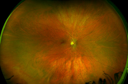
Retinal Tear Optos - Rhegmatogenous Retinal Detachment Widefield Optos Retina Image Bank / As brian had a history of retinal tears and the report of floaters seemed cause forconcern.
byAdmin-
0
Retinal Tear Optos - Rhegmatogenous Retinal Detachment Widefield Optos Retina Image Bank / As brian had a history of retinal tears and the report of floaters seemed cause forconcern.. An optomap® retinal exam provides: Today, millions of patients around the world have benefited from opto map® retinal imaging. The retina first tears, and then it detaches. The retina is the part of your eye that captures the image of what you are looking at, similar to film in a camera. Retinal holes and tears are commonly encountered during dilated fundus examination of both symptomatic and asymptomatic patients.
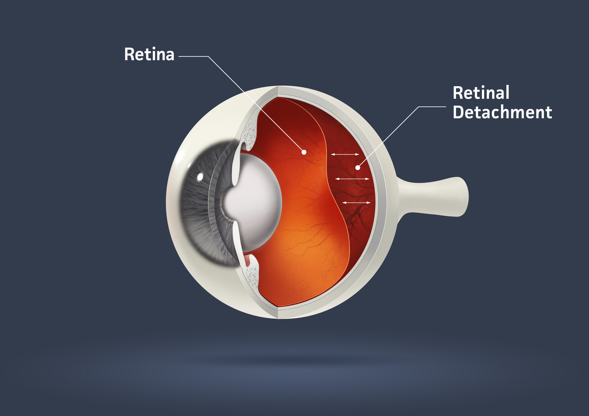
The retina is the part of your eye that captures the image of what you are looking at, similar to film in a camera. Operculated retinal tear an operculated tear results from vitreous traction that pulls a plug of sensory retina out into the vitreous cavity (see illustration). Another condition leading to retinal detachment is a giant retinal tear, defined as a retinal tear that extends 3 clock hours (90 degrees) or more around the circumference of the globe. Almost all clinically significant rds (greater than 2 disc diameters (dd) from the edge of the break or 1 dd posterior to the equator) if left untreated will eventually lead to a. Click on sample images to enlarge or download.
Retinal Holes And Tears Recognizing Pathology Optos from recognizingpathology.optos.com But, diseases such as macular degeneration, glaucoma, retinal tears or detachments, and other health problems such as diabetes and high blood pressure can be seen with a thorough exam of the retina. The retina is the part of your eye that captures the image of what you are looking at, similar to film in a camera. Another condition leading to retinal detachment is a giant retinal tear, defined as a retinal tear that extends 3 clock hours (90 degrees) or more around the circumference of the globe. Retinal tears when a retinal tear or hole hasn't yet progressed to detachment, your eye surgeon may suggest one of the following procedures to prevent retinal detachment and preserve vision. Gudeman was able to find a retinal tear and promptly refer them for surgery. We serve conshohocken, plymouth meeting, lafayette hill, and whitemarsh. Retinal detachment, optos, subretinal fluid, retinal tear photographer lauren r. The most frequent cause of an operculated tear is a pvd.
Today, millions of patients around the world have benefited from opto map® retinal imaging.
Optos retinal imaging the optos imaging technology allows us to give our patients a more thorough health evaluation. Retinal defects come in different shapes and sizes and may be either partial or full thickness. As brian had a history of retinal tears and the report of floaters seemed cause forconcern. Posterior uveitis can cause damage to the optic nerve, abnormally high pressure in the eye, cataracts or an accumulation of fluid within the retina which may cause retinal detachment. The most frequent cause of an operculated tear is a pvd. It shows the retina (where light and images hit), the optic disk (a spot on the retina that holds the optic nerve, which sends. Cancer can strike the eye just as it can strike almost any other part of the body. But, diseases such as macular degeneration, glaucoma, retinal tears or detachments, and other health problems such as diabetes and high blood pressure can be seen with a thorough exam of the retina. The device allows for the discovery of retinal melanomas and peripheral abnormalities such as retinal holes, retinal tears, and retinal detachments. Retinal holes and tears are commonly encountered during dilated fundus examination of both symptomatic and asymptomatic patients. This demonstrates that posterior vitreous detachment can produce. Retinal detachment, optos, subretinal fluid, retinal tear photographer lauren r. This is likely due to the vessels blocking the exiting light.
The superior steering revealed alarge pvd inferior and a possible retinal tear at eleveno'clock os. Almost all clinically significant rds (greater than 2 disc diameters (dd) from the edge of the break or 1 dd posterior to the equator) if left untreated will eventually lead to a. In many cases, this is a natural result of aging, but it. Cancer can strike the eye just as it can strike almost any other part of the body. Tearing of the retina may produce retinal or vitreous hemorrhages.
Optos Bennett Bloom Eye Centers from www.eyecenters.com An optomap® retinal exam provides: We offer the optomap® retinal exam as an important part of our eye exams. The device allows for the discovery of retinal melanomas and peripheral abnormalities such as retinal holes, retinal tears, and retinal detachments. Today, millions of patients around the world have benefited from opto map® retinal imaging. A retinal tear, or retinal detachment, occurs when the retina is torn from the underlying tissue. Retinal defects come in different shapes and sizes and may be either partial or full thickness. It is important to detect problems in the eye, like diabetes, macular degeneration, melanoma, bleeding, hypertensive retinopathy and glaucoma before they cause loss of vision. Retinal imaging takes a digital picture of the back of your eye.
There are many varieties of tears and breaks, but we can see them all with the optos and prescribe proper treatment accordingly.
Optomap retinal scan discovers critical retinal tear, saving vision and emphasizing the critical importance of optos' newly expanded line of optomap imaging devices lesley stabinsky compton wasn't having any vision problems when she went in for her annual optomap® eye exam with dr. These types of retinal detachments are the most common. Retinal detachment, optos, subretinal fluid, retinal tear photographer lauren r. It shows the retina (where light and images hit), the optic disk (a spot on the retina that holds the optic nerve, which sends. The neal eye group also offers an advanced photograph called an optos that can also help find serious retinal issues without the need for a dilated eye exam. But, diseases such as macular degeneration, glaucoma, retinal tears or detachments, and other health problems such as diabetes and high blood pressure can be seen with a thorough exam of the retina. The optomap® retinal exam produces an image that is as unique as you fingerprint and provides us with a wide view to look at the health of your retina. These are particularly clinically useful in eyes with retinal vascular disease. Doctor performed pneumatic retinopexy in office and patient recovering well. Gudeman was able to find a retinal tear and promptly refer them for surgery. It is important to detect problems in the eye, like diabetes, macular degeneration, melanoma, bleeding, hypertensive retinopathy and glaucoma before they cause loss of vision. Some times the retinal vessels on the detachment will be dark in appearance, as seen in a retinoschisis. If you think that you are suffering from a retinal tear, please call your optometrists at the vision center in fort worth and lubbock, tx.
The device allows for the discovery of retinal melanomas and peripheral abnormalities such as retinal holes, retinal tears, and retinal detachments. Tearing of the retina may produce retinal or vitreous hemorrhages. Optomap retinal scan discovers critical retinal tear, saving vision and emphasizing the critical importance of optos' newly expanded line of optomap imaging devices lesley stabinsky compton wasn't having any vision problems when she went in for her annual optomap® eye exam with dr. Another condition leading to retinal detachment is a giant retinal tear, defined as a retinal tear that extends 3 clock hours (90 degrees) or more around the circumference of the globe. As brian had a history of retinal tears and the report of floaters seemed cause forconcern.
The Top Signs You Have Retinal Detachment Drs Campbell Cunningham Taylor And Haun from www.ccteyes.com Often, these lesions are found in association with lattice degeneration. Operculated retinal tear an operculated tear results from vitreous traction that pulls a plug of sensory retina out into the vitreous cavity (see illustration). We serve conshohocken, plymouth meeting, lafayette hill, and whitemarsh. We offer the optomap® retinal exam as an important part of our eye exams. Tearing of the retina may produce retinal or vitreous hemorrhages. Gudeman was able to find a retinal tear and promptly refer them for surgery. Decided to take an optos image and this is what we found. Optos retinal imaging the optos imaging technology allows us to give our patients a more thorough health evaluation.
This demonstrates that posterior vitreous detachment can produce.
The most frequent cause of an operculated tear is a pvd. Today, millions of patients around the world have benefited from opto map® retinal imaging. The device allows for the discovery of retinal melanomas and peripheral abnormalities such as retinal holes, retinal tears, and retinal detachments. There are many varieties of tears and breaks, but we can see them all with the optos and prescribe proper treatment accordingly. The optomap® retinal exam produces an image that is as unique as you fingerprint and provides us with a wide view to look at the health of your retina. We offer the optomap® retinal exam as an important part of our eye exams. Tearing of the retina may produce retinal or vitreous hemorrhages. The retina is the part of your eye that captures the image of what you are looking at, similar to film in a camera. We serve conshohocken, plymouth meeting, lafayette hill, and whitemarsh. Cancer can strike the eye just as it can strike almost any other part of the body. In many cases, this is a natural result of aging, but it. Gudeman was able to find a retinal tear and promptly refer them for surgery. Whaley imaging device optos description noticed an inferior visual field defect on a patient with history of vitreous hemorrhage.
Cancer can strike the eye just as it can strike almost any other part of the body retinal tear. The most common reason for a rd is a retinal tear and the most common reason for a tear is a pvd.